2025: Volume 4, Issue 2
Past Issues
 Abstract
  PDF
PDFCase Report of a Congenital Anterior Urethral Diverticulum in a Sudanese Child
Muawia Ahmed Hassan1, Adil Ibrahim2, Mohamed Elimam Mohamed Ahmed3,*
1Associate Professor of Urology, University of AL Neelain, Sudan
2Associate Professor of Urology, University of Khartoum, Sudan
3Professor of Urology, University of Gezira, Sudan
*Corresponding author: Mohamed Elimam Mohamed Ahmed, MD, FRCS, Professor of Urology, University of Gezira, Sudan, Phone: 00249912362293, E-mail: [email protected]
Received Date: July 20, 2025
Publication Date: September 09, 2025
Citation: Hassan MA, et al. (2025). Case Report of a Congenital Anterior Urethral Diverticulum in a Sudanese Child. Cases. 4(2):23.
Copyright: Hassan MA, et al. © (2025).
ABSTRACT
Congenital anterior urethral diverticulum (CAUD) is an extremely rare. Here, we describe our experience with a 10-year-old boy. We describe an ambiguous clinical course of the disease. Investigations, management courses, and follow-up are described in detail, along with an insight into the literature.
Keywords: Urethra, Diverticulum, Sudan
INTRODUCTION
Congenital urethral diverticulum (CUD) is a rare urological anomaly characterized by an outpouching or saccular dilatation of the urethra, present at birth. While urethral diverticula can be acquired due to various factors such as infection, trauma, or iatrogenic injury, the congenital form represents a distinct clinical entity, primarily affecting male children [1,2]. The incidence of CUD is low, making it an uncommon cause of lower urinary tract obstruction in paediatric populations [3].
The embryological basis of CUD remains a subject of ongoing debate, with several hypotheses proposed. These include a developmental defect in the corpus spongiosum, cystic dilatation of periurethral glands, or sequestration of epithelial nests during urethral fold closure [1,4].
Regardless of its precise etiology, the presence of a CUD can lead to a spectrum of clinical manifestations, ranging from asymptomatic cases to severe obstructive uropathy. Common presentations in children include a poor urinary stream, post-void dribbling, recurrent urinary tract infections, and a palpable swelling on the ventral aspect of the penis, which may enlarge during micturition [1,5]. In some instances, CUD can be associated with other congenital anomalies, such as posterior urethral valves, further complicating diagnosis and management [6].
Early and accurate diagnosis is crucial to prevent long-term complications, including bladder dysfunction, hydronephrosis, and renal impairment [6]. Diagnostic modalities typically involve a combination of clinical examination and imaging studies. Voiding cystourethrography (VCUG) and retrograde urethrography are considered cornerstone investigations, often revealing the characteristic diverticular sac and its communication with the urethra [1,5]. Ultrasonography can complement these studies by assessing changes in the upper urinary tract and bladder wall. At the same time, magnetic resonance imaging (MRI) offers detailed anatomical visualization, which is beneficial for surgical planning [7].
Management of CUD is primarily surgical, with the approach tailored to the diverticulum's size, location, and the degree of associated obstruction. Treatment options range from endoscopic procedures, such as transurethral resection of the distal obstructing lip, to open diverticulectomy with urethroplasty for larger or more complex cases [1,3]. Surgical intervention aims to relieve obstruction, prevent urinary stasis, and preserve renal function. This introduction aims to provide a comprehensive overview of congenital urethral diverticulum, highlighting its clinical significance, diagnostic challenges, and therapeutic considerations.
THE CASE REPORT
Our patient is a 10-year-old male originally from West Darfur. He presented with difficulty urinating a few months before visiting our department at Sharg Alneel Model Hospital.
Previously, he had been seen by a physician in a nearby district, who diagnosed him with a urethral stone. According to the patient's family, the doctor performed an incision in the middle of the scrotum to remove the stone. A urinary catheter was left in place for one week and was subsequently removed, with the wound healing satisfactorily and without any complications such as fistulas. Unfortunately, the patient's urinary stream did not improve. He continued to experience repeated urinary tract infections, dribbling, and severe pain during urination.
Interestingly, a swelling was observed that increased markedly during urination but gradually decreased in size, though it did not disappear entirely. There was no history of trauma. A systematic review of the patient's medical history was unremarkable, except for the previous surgery. The patient had no known allergies to medications. Additionally, there was no family history of similar conditions. Socially, he is an orphan who lost his father at an early age, and his uncle is currently caring for him. The family comes from a destitute background with limited socioeconomic resources.
During the examination, the patient appeared generally well but was in pain. He was neither pale nor jaundiced and had no signs of cyanosis. A corneal opacity was noted. The examinations of the cardiovascular, respiratory, and CNS systems were normal. A local examination revealed a significant scrotal swelling approximately the size of an orange, with an unabsorbed Vicryl stitch from the previous surgery. The swelling was neither hot nor tender and had a cystic consistency. No other abnormalities were detected.
Laboratory investigations showed the following:
Urine general pus cells 18-20/HF RBCs 3-4/HPF
Serum creatinine 1mg/dl. Blood urea 25mg/dl, Hb 11gm/dl TWBC 6000.
An abdominal ultrasound revealed that the left kidney is of normal size with no signs of hydronephrosis. In contrast, the right kidney is smaller and contains stones at the lower pole. The urinary bladder appears normal, but the right testis is not visualized in the scrotum or the inguinal canal.
A CT tomogram identified a large obstructing stone in the right lower ureter. Subsequently, a urethrogram (MCUG) was performed, which showed a significant urethral diverticulum (Figure 2).
Management
Open urethrolithotomy was performed, resulting in an uneventful postoperative course. A temporary suprapubic cystostomy was also conducted (Figure 1). One month later, we proceeded with the operation on the diverticulum. Prior to the surgery, a cystoscopy was performed, which revealed a wide cavity in the peno-bulbar region.
Under general anesthesia, a perineal incision with scrotal extension was made. The large anterior urethral diverticulum was completely dissected and excised Figures 3 & 4), allowing for the tailoring of an adequate urethral lumen. Urethral reconstruction was carried out over a 10 Fr Foley catheter. The postoperative period was uneventful, and follow-up cystoscopy conducted one month later showed a normal urethral lumen with no valves present in the urethra. The urethral catheter was subsequently removed.
There was a marked improvement in urinary flow, and the child was able to void comfortably. The suprapubic catheter was removed a few days after clamping, and the suprapubic wound healed satisfactorily. Unfortunately, we lost contact with the patient after this.
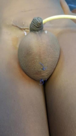
Figure 1. Suprapubic catheterization before surgery.
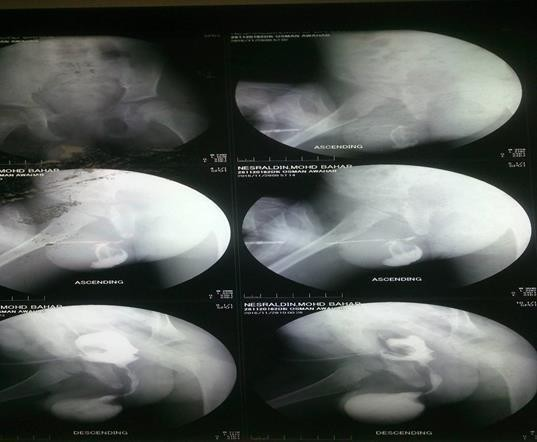
Figure 2. MCUG of the diverticulum.
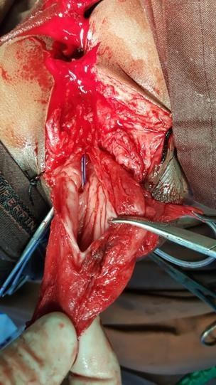
Figure 3. Excision of the diverticulum.
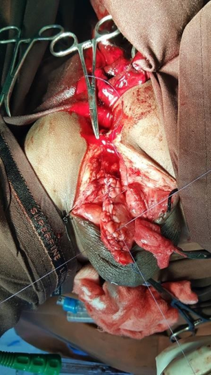
Figure 4. Excision of the diverticulum.
DISCUSSION
Congenital anterior urethral diverticulum (CAUD) is an exceptionally rare urological anomaly [8]. Paulhac et al. (2003) reviewed existing literature, identifying only 250 reported cases by 2003, highlighting its infrequent occurrence [9]. CAUD is estimated to account for 10–20% of all urethral diverticula [10]. It can manifest as an isolated condition or in conjunction with urethral valves, with some theories suggesting that CAUD may arise as a consequence of urethral valves [8].
Various theories have been proposed to elucidate the etiopathogenesis of CAUD. These include developmental defects in the corpus spongiosum, cystic dilatation of urethral glands, and sequestration of urethral anlage following incomplete fusion of urethral folds [11]. Rawat et al. (2012) described six patients presenting with anterior urethral valves, all of whom also had associated diverticula [12]. This observation supports the hypothesis that urethral valves may lead to urinary flow obstruction, subsequently promoting diverticular formation. Conversely, McLellan et al. (1995) postulated that rupture of Cowper's glands could result in a cyst (syringocele), with the distal lip of the cyst potentially acting as an obstructing valve leaflet and leading to subsequent diverticulum formation [13]. In the present case, preoperative and postoperative cystoscopic examinations revealed no evidence of urethral valves.
CLINICAL PRESENTATION
CAUD typically presents with obstructive lower urinary tract symptoms, including a weak urinary stream, post-micturition dribbling, and dysuria [14]. The patient in this report experienced severe dysuria, characterized by distress during micturition.
In rare instances, CAUD may be associated with urolithiasis [15]. These calculi can occasionally attain substantial sizes. The precise method by which the initial diagnosis of a stone was made in this patient's case remains unclear. It is hypothesized that the treating physician, operating in a peripheral hospital with limited imaging capabilities, may have palpated a scrotal stone.
Shuzhu et al. (2006) reported a case of CAUD associated with primary infertility in an adult male in his fourth decade [16].
Age at Presentation
CAUD can manifest at any age, ranging from infancy to adulthood [8]. Antenatal diagnosis is also possible; for instance, Coplen and Austin (2003) documented three cases diagnosed in utero and managed during the early postnatal period [17].
Location
In the current case, the diverticulum was situated in the peno-bulbar segment of the urethra, which is consistent with the majority of reported cases. However, CAUD can occur in any part of the anterior urethra. Kadian et al. (2009) reported a case located in the subcoronal portion of the penile urethra [18].
Imaging
Retrograde or micturating cystourethrography remains the gold standard for diagnosing CAUD. Goyal et al. (2003) described a novel ultrasound-based technique, demonstrating its utility as an alternative to contrast studies [19]. More recently, magnetic resonance imaging (MRI) has been incorporated into the diagnostic armamentarium [20].
Management
In the reported case, the initial priority was the removal of a ureteral stone, which may have developed as a sequela of recurrent sepsis. Given the high prevalence of stone disease in the patient's geographical area, a suprapubic catheter was concurrently placed for urinary diversion. The diverticulectomy was performed one month later.
The management of CAUD is largely dictated by its size. Small diverticula can often be managed endoscopically, whereas larger diverticula typically necessitate surgical excision and urethral reconstruction [21,22]. Adhering to this principle, the diverticulum in this patient was excised, followed by urethral reconstruction. Minimal adhesions were encountered despite prior intervention.
CONCLUSION
CAUD is an exceedingly rare condition. The presented case followed a classic clinical course. Notably, the inadvertent intervention by a district physician modified the diagnostic and management trajectory. Furthermore, this case was unique due to its association with ureteral and renal stone disease, and a corneal opacity that unfortunately could not be investigated due to loss of patient follow-up.
ACKNOWLEDGEMENTS
None.
CONFLICT OF INTEREST DECLARATION
The authors declare that they have no affiliations with or involvement in any organization or entity with any financial interest in the subject matter or materials discussed in this manuscript.
REFERENCES
- Okur M, Aydogdu B, Ciftci E, Arslan M, Yildiz M. (2015). A congenital anterior urethral diverticulum associated with obstructive urinary symptoms in a 1 -year-old male child: A case report and review of the literature. Pediatr Urol Case Rep. 2(3):25 -30.
- Dangol E, Thapa B, Thapa AB. (2024). Congenital Anterior Urethral Diverticulum: A Case Report. NMJ. 6(1):674-676.
- Önal O, Caylan AE, Uçar M. (2024). A Rare Case in Pediatric Urology: Coexistence of Congenital Anterior Urethral Diverticulum and Posterior Urethral Valve. J Urol Surg. 11(4):231-234.
- Smith GHH, Deshpande A, Tang RWK. (2014). Uncommon causes of anterior urethral diverticula in children: Two cases and review of literature. Urol Ann. 6(2):166-169.
- Singh A, Singh S, Singh M. (2014). Congenital anterior urethral diverticulum: A case report. J Pediatr Urol. 10(6):1108-1110.
- Gebreselassie HA, Girma H, Temesgen F. (2021). Congenital anterior urethral diverticula with posterior urethral valve: A rare combination, case report. Urol Case Rep. 35:101517.
- Chung BH, Lee KS, Kim YJ, Lee JW, Kim TH. (2008). Pre- and Postoperative Evaluation of Urethral Diverticulum. AJR Am J Roentgenol. 190(5):1353-1360.
- Eshiba A, Ashour K, Zein M. (2011). Congenital anterior urethral diverticulum. Pediatr Surg Int. 27(1):97-100.
- Paulhac P, Fourcade L, Lesaux N, Alain JL, Colombeau P. (2003). Anterior urethral valves and diverticula. BJU Int. 92(5):506-509.
- Bayarktar H. (2010). Urethral Diverticula. Urology. 76(2):498-502.
- Singh SK, Ansari MS. (2012). Congenital anterior urethral diverticulum. J Indian Assoc Pediatr Surg. 17(4):175-177.
- Rawat J, Khan TR, Singh S, Maletha M, Kureel S. (2012). Congenital anterior urethral valves and diverticula: Diagnosis and management in six cases. J Pediatr Urol. 8(1):21-25.
- McLellan DL, Gaston MV, Diamond DA, Lebowitz RL, Mandell J, Atala A, et al. (1995). Anterior urethral valves and diverticula in children: a result of ruptured Cowper's duct cyst? Urology. 46(3):395-398.
- Dewan PA. (2006). Anterior urethral diverticulum. BJU Int. 97(4):861-864.
- Rajendiran S, Bhatnagar V, Lal R, Gupta AK, Mitra DK. (2000). Anterior urethral diverticulum with calculus. Pediatr Surg Int. 16(5-6):436-437.
- Shuzhu C, Min W, Weijing Y, Yidong L. (2006). Congenital urethral diverticulum with infertility in an adult man and review of the literature. Int J Urol. 13(4):450-452.
- Coplen DE, Austin PF. (2003). Prenatal diagnosis and neonatal management of congenital urethral diverticulum. J Urol. 169(4):1538-1540.
- Kadian YS, Rattan KN, Singh M, Kajal P. (2009). Congenital anterior urethral diverticulum in children: a case report and review. J Neonatal Surg. 1(3):31.
- Goyal M, Sharma R, Gupta DK, Sharma A, Berry M. (2003). Congenital anterior urethral diverticulum: sonographic diagnosis. J Ultrasound Med. 22(12):1379-1382.
- Chan KW, Chu WH. (2012). Giant urethral diverticulum calculus presenting as scrotal abscess. Hong Kong Med J. 18(1):66-67.
- Glassberg KI, LaMarca A. (2005). Congenital anterior urethral diverticulum. J Pediatr Urol. 1(1):2-7.
- Kajbafzadeh AM, Sina A, Tourchi A. (2011). Management of large congenital anterior urethral diverticula. J Urol. 185(6 Suppl):2548-2553.