2025: Volume 4, Issue 1
Past Issues
 Abstract
  PDF
PDFGiant Cell Tumour with Two Lesions- Case Report
Arshika Faisal, Maryum Anwar, Asna Shabbir, Saleha Haider, Ramna Shafique*
Ziauddin Medical College, Ziauddin Medical University, Karachi, Pakistan
*Corresponding author: Miss. Ramna Shafique, Final-year Medical Student, Ziauddin Medical College, Ziauddin Medical University, B Shahrah-e-Ghalib Rd, Block 6 Clifton, 75000, Karachi, Pakistan, Tel: 03493659018, E-mail: [email protected]
Received Date: March 04, 2025
Publication Date: April 09, 2025
Citation: Faisal A, et al. (2025). Giant Cell Tumour with Two Lesions- Case Report. Cases. 4(1):21.
Copyright: Faisal A, et al. © (2025).
ABSTRACT
Giant cell tumour (GCT) is one of the most common benign bone tumours, affecting the age group of 20 to 40 years. The epiphysis or metaphysis of long bones is most commonly affected. The author reports the case of a 15-year-old Southeast Asian who presented with pain and swelling in the left distal tibia. Upon doing an investigation and biopsy, a giant cell tumour with multiple lesions was identified in the left distal tibia, which was successfully treated.
Keywords: Multiple Lesions, Pain, Swelling, Tumour, Distal Tibia, Benign
INTRODUCTION
The giant cell tumor (GCT), a benign primary tumor of the bone, was initially reported by Cooper and Travers in 1818. One of the most prevalent benign bone tumors, giant cell tumors often affect young individuals between the ages of 20 and 40 [1]. About 20% of benign tumors examined by biopsy are giant cell tumors (GCTs), which account for 5% of all primary bone cancers [2,3]. More than half of these lesions appear in the third and fourth decades of life [1]. About 85-90% of GCT occurs in the meta-epiphyseal area of long bones, with the knee accounting for 50%, followed by the distal radius, sacrum, axial skeleton, and tiny bones of the hands and feet [3-5]. Compared to groups in the West, Asians have strangely a much greater prevalence of it. Although GCT may affect all races, this has not been explained to date [1]. Despite the fact that GCT-TS can occur at any age, it often manifests between the ages of 30 and 50, with a 2:1 female preponderance [6].
Clinical symptoms are nonspecific and may include local pain and swelling [3]. The only curative treatment for giant cell tumor is adequate surgery [2]; options are intralesional curettage, intralesional curettage and bone grafting, intralesional curettage and insertion of polymethylmethacrylate (PMMA), primary resection, radiation therapy, and embolization of the feeding vessels [2]. Various local adjuvants such as cryosurgery, phenol, bone cement, zoledronic acid, hydrogen peroxide (H?O?), and argon beam, as well as systemic treatments such as bisphosphonates, interferon alpha (IFN-α), and denosumab, have been reported to reduce the risk for local recurrence, with varying outcomes, function, and complications for the patients [7,8].
CASE PRESENTATION
A 15-year-old female from Baluchistan, Pakistan, presented with a 2-year history of pain and swelling at the left medial ankle, which initiated after a history of trauma with ankle dislocation.
On general physical examination, the patient was responsive but unable to walk without support, and her movements were limited in the left leg. Her vitals were BP: 116/63 mmHg, pulse: 74 beats per minute, respiratory rate: 20 breaths per minute, temperature: 96 Fahrenheit, O? saturation: 98% at room air. Musculoskeletal examination revealed red, oedematous swelling on the medial aspect of the left ankle. The central nervous system was intact with a Glasgow Coma Scale of 15/15. Examination of the abdomen and cardiovascular system did not reveal any abnormalities. There was no history of fever or weight loss.
Clinical investigation was non-significant (Table 1). Upon radiological imaging, X-ray (Image 1) revealed two well-defined, expansile, eccentric, lucent lesions with internal septation in the radial epiphysis of the left distal tibia, most likely neoplastic lesions. Mild soft tissue swelling was also seen. Normal joint space with no gross evidence of acute displaced fracture or dislocation. There was no significant family history of malignancy or neoplastic tumours.
Table 1. Lab Investigation
|
HBsAg |
Negative |
|
Anti HCV |
Negative |
|
Hb |
12.3 gm/dL |
|
HTC |
39% |
|
MCV |
84fL |
|
Total WBC Count |
8.1 x 10E9/L |
|
Neutrophils |
61% |
|
Lymphocytes |
32% |
|
Monocytes |
5% |
|
Eosinophils |
2% |
|
Platelet Count |
376 x 10E9/L |
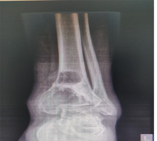
Image 1. Ankle X-ray of 15-year-old female, AP/LAT(LT). revealed two well defined eccentric, expansile lucent lesions in radial epiphysis.
Based on the clinical and radiological findings, a diagnosis of giant cell tumour was suspected. An incisional biopsy was performed, and the histopathological examination showed multiple tan-white irregular soft tissue pieces that measure 7 x 7.5 x 2.5 cm in aggregate. Sections examined revealed bony trabeculae and connective tissue exhibiting a lesion composed of scattered clusters of multinucleated osteoclastic giant cells. The surrounding background was hemorrhagic. Immunohistochemical stain H3G34W was positive. Upon confirmation of the diagnosis, the patient was scheduled for surgical resection of the bone. Excision of the giant cell tumour with bone cementing and bone grafting was done under general anesthesia. An incision was given at the distal tibia anteriorly. The tumour cavity was curetted and excised along with the necrotic tissue, followed by another biopsy. Bone graft taken from the left iliac crest was grafted inside the hollow distal tibia along with bone cement, followed by Dynacast short leg application. The patient showed satisfactory recovery and was advised for no weight bearing for 15 days and complete weight bearing without support after 60 days, followed with physical therapy. Regular follow-up visits were scheduled to monitor the patient’s progress.
DISCUSSION
In 1818, Cooper and Travers published the first account of the giant cell tumour of the bone. Its potential for malignancy was highlighted by Virchow, while Nelaton emphasised its local aggression [9].
Giant cell tumour (GCT), a commonly occurring benign bone tumour in young adults (20–40 years), has a significant rate of recurrence and the potential to become aggressive [1]. Giant cell tumours of the bone usually affect skeletally mature people, with the highest incidence in the third decade of life, and have a pronounced female preponderance [10]. Although primary and recurrent giant cell tumours of the bone are typically benign, they can occasionally transform into malignant tumours. Giant cell bone tumours can develop malignancies that are either primary (near a benign giant cell bone tumour at the time of the initial diagnosis) or secondary (at the site of a giant cell bone tumour that has already undergone treatment) [11]. GCTs are relatively common, accounting for 5% of all tumours, 20% of benign bone tumours, and 20% of all tumours [1]. In the Asian population, GCTs make up 18% of all non-haematogenous primary bone tumours, as reported in a recent study. The incidence of GCT in the immature skeleton is only mentioned in Western literature and ranges from 1.8% to 10.6%. According to Schutte and Taconis, the epiphysis is never involved in tubular bones with open epiphyseal growth plates, and epiphyseal involvement increases with advancing age. The most frequent site, according to a study done in India, was near the knee, with 9 of the 17 lesions (or 53%) occurring in the lower end of the femur and the upper end of the tibia [10]. The average local recurrence rate is 33%, with a range of 20% to 50% [1].
They typically present as solitary lesions. The multicenter occurrence of a GCT is extremely rare, with only around 1-2% of cases described in the literature [12]. In our case, however, the patient presented with two well-defined expansile and eccentric lesions on her left medial ankle, revealed in X-ray imaging. It frequently manifests in the long bone meta-epiphysis and typically affects the subchondral bone without impacting the articular surface. Larger tumours, however, can occasionally invade the metaphysis and, much less frequently, the diaphysis [13].
Histologically, the lesions appear cellular with a background network of mononuclear stromal cells, with multinucleated giant cells being the characteristic cell type. The cells may be spindle-shaped, oval-shaped, or plump with prominent mitotic activity, and rarely, cellular atypia [14]. Similarly, in our case, histopathological examination revealed a lesion primarily composed of scattered clusters of spindles to oval-shaped, multinucleated osteoclastic giant cells with a hemorrhagic background but absent mitotic activity and cellular pleomorphism. Imaging techniques like computed tomography (CT) scans and magnetic resonance imaging (MRI) may be essential to confirm the typical subchondral location of these lesions within the bone itself and the extent of a soft tissue mass, either beyond the bone cortex or through the nearby joint [1].
The differential diagnosis based on the radiographic findings can include lytic metastatic lesions (particularly a vascular metastasis from thyroid or renal cell carcinoma). Primary bone tumour, Brown tumour of hyperparathyroidism, non-ossifying fibroma, Aneurysmal bone cyst, Fibrous metaphyseal defects, Osteoblastoma, chondroblastoma, malignant fibrous histiocytoma, telangiectatic osteosarcoma [1].
Despite being an uncommon, mostly benign tumour, it may act in an unexpected way regardless of the findings of radiological tests. Hence, results of the radiographic examination can assist in reaching a differential diagnosis; they cannot be conclusive. The gold standard for diagnosis continues to be histological analysis [10].
According to Campanacci et al., GCTs can be classified into three grades based on their radiographic appearance: grade 1 lesions (latent) have an intact cortex and well-defined margins; grade 2 lesions (active) have a relatively well-defined margin without a radiopaque rim, with a thinned and moderately expanded cortex; and grade 3 lesions (aggressive) have indistinct borders and cortical destruction. In a select number of patients with Campanacci grade 3 lesions that have joint invasion, a multiplanar soft tissue component, and when the tumour covers a large amount of subchondral bone, wide excision and reconstruction is the choice of treatment [15].The main objectives of the treatment and management of a GCT in the paediatric age group are local control, maintaining joint function, and preserving the physis, although the increased likelihood of the tumour’s recurrence is a primary concern as well [16]. There are several treatment modalities for giant cell tumours that have been described in the literature previously. Amputations, wide resections, or reconstructions were once used to treat GCT. However, because GCT is a benign but regionally aggressive tumour, a local intralesional surgical approach is usually appropriate. The recommended treatments include curettage, curettage and bone grafting, curettage and polymethylmethacrylate (PMMA) insertion, and primary resection. For pelvic and sacral tumours that are inoperable, radiation therapy and feeding vessel embolisation are used [1].
According to Schajowicz [17], when paired with adjuvant therapy, curettage, an inadequate oncological technique when used alone, has a more effective overall outcome than one-block excision, especially in terms of functioning. Some adjuvant therapies include phenol, liquid nitrogen, or hydrogen peroxide and coagulation using argon beam, and each of these treatment modalities has its own advantages and disadvantages [18].
Despite the fact that they are rarely lethal, benign bone tumours may cause a significant disruption in the local bony architecture, which can be particularly problematic in periarticular regions [18]. According to a study published by Hosseinzadeh S et al., giant cell tumours of the bone may be complicated by tumour recurrence, osteoarthritis of the knee joint, stress fracture, limited movement, pulmonary metastasis, local and deep infections, osteomyelitis, joint degeneration, and hardware failure [1].
CONCLUSION
The majority of the reported cases of GCT of the bone that have been documented in literature present with solitary lesions. In our case, however, the 15-year-old presented with two lytic lesions on her distal tibia, which is a rare finding. Moreover, histopathological examination is the most crucial diagnostic tool for GCT of the bone, with prompt diagnosis and effective treatment leading to a positive result. Currently, the patient at 1-month follow-up is doing well and walking with support, comfortably without any pain. Her wound dressings are being changed at regular intervals, and there are no signs of recurrences.
ACKNOWLEDGMENT
None.
CONFLICT OF INTEREST
The authors declared no conflict of interest.
REFERENCES
- Hosseinzadeh S, De Jesus O. (2023). Giant Cell Tumor. StatPearls. Available at: https://www.ncbi.nlm.nih.gov/books/NBK559229/
- Sobti A, Agrawal P, Agarwala S, Agarwal M. (2016). Giant Cell Tumor of Bone - An Overview. Arch Bone Jt Surg. 4(1):2-9.
- Tejwani SG, Hame SL, Eckardt JJ. (2004). Subchondral giant-cell tumor of the proximal tibia: arthroscopic treatment for accelerated articular cartilage and meniscal degeneration in two patients. Arthroscopy. 20(6):644-649.
- van der Heijden L, Dijkstra PD, van de Sande MA, Kroep JR, Nout RA, van Rijswijk CS, et al. (2014). The clinical approach toward giant cell tumor of bone. Oncologist. 19(5):550-561.
- Siddiqui YS, Zahid M, Bin Sabir A; Julfiqar. (2011). Giant cell tumor of the first metatarsal. J Cancer Res Ther. 7(2):208-210.
- Reilly KE, Stern PJ, Dale JA. (1999). Recurrent giant cell tumors of the tendon sheath. J Hand Surg Am. 24(6):1298-1302.
- Ruggieri P, Mavrogenis AF, Ussia G, Angelini A, Papagelopoulos PJ, Mercuri M. (2010). Recurrence after and complications associated with adjuvant treatments for sacral giant cell tumor. Clin Orthop Relat Res. 468(11):2954-2961.
- Chawla S, Henshaw R, Seeger L, Choy E, Blay JY, Ferrari S, et al. (2013). Safety and efficacy of denosumab for adults and skeletally mature adolescents with giant cell tumour of bone: interim analysis of an open-label, parallel-group, phase 2 study. Lancet Oncol. 14(9):901-908.
- Vanni D, Pantalone A, Andreoli E, Caldora P, Salini V. (2012). Giant cell tumor of the distal ulna: a case report. J Med Case Rep. 6:143.
- Puri A, Agarwal MG, Shah M, Jambhekar NA, Anchan C, Behle S. (2007). Giant cell tumor of bone in children and adolescents. J Pediatr Orthop. 27(6):635-639.
- Palmerini E, Picci P, Reichardt P, Downey G. (2019). Malignancy in Giant Cell Tumor of Bone: A Review of the Literature. Technol Cancer Res Treat. 18:1533033819840000.
- Hoch B, Inwards C, Sundaram M, Rosenberg AE. (2006). Multicentric giant cell tumor of bone. Clinicopathologic analysis of thirty cases. J Bone Joint Surg Am. 88(9):1998-2008.
- Vanni D, Pantalone A, Andreoli E, Caldora P, Salini V. (2012). Giant cell tumor of the distal ulna: a case report. J Med Case Rep. 6:143.
- Steensma MR, Tyler WK, Shaber AG, Goldring SR, Ross FP, Williams BO, et al. (2013). Targeting the giant cell tumor stromal cell: functional characterization and a novel therapeutic strategy. PLoS One. 8(7):e69101.
- Campanacci M, Baldini N, Boriani S, Sudanese A. (1987). Giant-cell tumor of bone. J Bone Joint Surg Am. 69(1):106-114.
- Sarkar S, Laik JK, Kaushal R, Mishra M, Rajak M. (2023). A Rare Giant Cell Tumour in the Distal Radius of a Seven-Year-Old Girl: A Case Report. Cureus. 15(6):e40270.
- Schajowicz F. (1994). Tumors and Tumor-like Lesions of Bone: Pathology, Radiology and Treatment. Germany: Springer-Verlag.
- Balke M, Schremper L, Gebert C, Ahrens H, Streitbuerger A, Koehler G, et al. (2008). Giant cell tumor of bone: treatment and outcome of 214 cases. J Cancer Res Clin Oncol. 134(9):969-978.