2025: Volume 4, Issue 1
Past Issues
 Abstract
  PDF
PDFA Novel Technique for Iliac Crest Reconstruction After Autologous Bone Harvest with Titanium Bone Plate and Screws, And HydroSetTM - Case Report
Eber Luis de Lima Steolo1,*, Michael Wirey2, Brett Barnett3, Paul Sata Kamash4
1Doctor of Dental Surgery, Oral and Maxillofacial surgeon, PhD in OMFS, Post-Doctorate at Baylor University Medical Center and Baylor College of Dentistry, Dallas - TX, USA, Assistant Professor OMFS Department at West Virginia University School of Dentistry (WVVUSOD), Morgantown - WV, Active member of the ASTMJS, USA
2Doctor of Dental Surgery, Fourth Year Resident – Resident Chief - OMFS Department at WVUSOD, Morgantown-WV, USA
3Doctor of Dental Surgery, Third Year Resident OMFS Department at WVUSOD, Morgantown-WV, USA
4Doctor of Dental Surgery, Second Year Resident OMFS Department at WVUSOD, Morgantown-WV, USA
*Corresponding author: Eber Luis de Lima Steolo, DDS, EdS, PhD, Doctor of Dental Surgery, Oral and Maxillofacial surgeon, PhD in OMFS, Post-Doctorate at Baylor University Medical Center and Baylor College of Dentistry, Dallas - TX, USA, Assistant Professor OMFS Department at West Virginia University School of Dentistry (WVVUSOD), Morgantown - WV, Active member of the ASTMJS, USA, Phone: 304 2828438, E-mails: [email protected]; [email protected]
Received Date: January 31, 2025
Publication Date: March 21, 2025
Citation: Steolo ELDL, et al. (2025). A Novel Technique for Iliac Crest Reconstruction After Autologous Bone Harvest with Titanium Bone Plate and Screws, And HydroSetTM - Case Report. Cases. 4(1):20.
Copyright: Steolo ELDL, et al. © (2025).
ABSTRACT
Background: The iliac crest (IC) has been for decades the golden standard autologous bone graft donor site, although associated with pain, intense intraoperative cancellous bone bleeding, postoperative hematoma formation, hip aesthetic defect, iliac instability and/or fracture. Reconstruction of the IC after bone grafting has recent gained attention minimizing complications, bringing this IC to the main option when autogenous bone grafting is needed in great quantity. Methods: The authors present a case report on a patient who underwent inner anterior left IC corticocancellous harvesting for maxillary bone reconstruction. Rigid fixation of the defect was obtained with a 2.0 titanium bone plate fixated to the superior border of the IC with four titanium screws, and a self-setting calcium phosphate cement intended for use in the repair or restoration of bony contour in human skeleton. Results: Trans-operative bleeding was minimal due to a careful use of ultrasonic surgery technique, reducing postoperative hematoma formation. Minimal postoperative donor site pain and early painless ambulation on this patient significantly differed from previous descriptions on scientific literature due to several aspects described in details on this article but the authors impute the final success of this new surgical technique to a very careful IC muscle dissection and primary stability of the hip with titanium bone plating and screw fixation, and hydroxyapatite cement extra support. Conclusion: Even though there is a tremendous stigma when iliac crest bone harvesting (ICBH) is the option for autologous corticocancellous bone harvesting due to several aspects such as bleeding, pain, hematoma, ambulation and pronounced scar. A careful and punctilious surgical technique which avoids drills and saws, followed by reconstruction of the IC with rigid fixation, mediated by a meticulous postoperative pain protocol, intraoperative bleeding can be attenuated, postoperative pain tremendously reduced, early ambulation staggeringly improved, and scar reduced with appropriate and compressive postoperative dressings. The surgical procedures described on this article should be considered as an excellent technique for ICBH due to its various benefits when compared to the traditional techniques which do not consider donor site reconstruction.
Keywords: Iliac Crest Bone Graft, Bone Harvest Morbidity, Reconstruction of Pelvic Donor Site, Bone Cement, Hydroxyapatite Iliac Reconstruction, Autologous Bone Harvest
INTRODUCTION
Autologous bone grafts are widely used in clinical orthopedics due to their biological and non-immunologic properties compared to other materials [1]. And out of autologous bone grafts, the IC is a preferred donor site due to its capacity to yield good amount of cortical and generous quantity of cancellous bone for grafting purposes, straightforward accessibility, low risk of complications and/or morbidity, and it is considered the gold standard of donor sites [2,3].
Autologous iliac bone demonstrates optimal osteogenic, osteoinductive, and osteoconductive properties [4], and it can be utilized in numerous Oral and Maxillofacial procedures such as sinus lifting, ridge augmentation, and defect reconstruction [5].
Research comparing iliac bone grafts with other materials such as allografts, xenografts, and synthetic substitutes demonstrate superior integration and long-term stability [6,7]. However, these benefits are often weighed against the potential for donor site complications.
Nevertheless, complication rates following ICBH have been reported from 2% to 49%. Other complications of this procedure include pain, secondary fracture, ipsilateral thigh superficial numbness, infection, abdominal hernia, gait disturbances, poor cosmetic appearance, and depression of the surgical area [8-10,2]. Patients suffering from pain in the donor site refer it has an effect on sleep within one month after surgery, and even 13% to 20% of these patients end up experiencing chronic pain [11,12]. In addition, harvesting a large graft from IC bone leads to some problems such as depression of the surgical area, poor cosmetic appearance, having influence on walking, recreation, and household chores [11].
Reconstruction of the IC after bone harvesting is a growing area of interest due to the desire to minimize donor site complications and maintain bone structural integrity. Several investigative scientific studies suggest that reconstruction of the donor site provides reduction in postoperative pain [13], improvement in local aesthetics and function outcomes [14], along with preventing long-term complications such as herniation or pelvic instability [2].
Some studies have indicated that reconstruction of IC defects after harvesting can reduce several complications [13-18].
Techniques in reconstructing the IC include several materials such as: a) hydroxyapatite-calcium triphosphate biphasic compound [19], b) autologous bone [15], c) bovine cancellous grafts [17], d) polymethyl methacrylate bone cement [20], e) allografts fixed with cannulated screws [18], and f) bone cement with cancellous screws [2].
In this case report, the authors describe a new surgical technique using titanium (Ti) bone plate and screws, along with hydroxyapatite bone cement (HABC) to repair the iliac monocortical bone defect after autologous harvesting for maxillary reconstruction.
The purpose of this study was to describe and evaluate the benefits of this new approach developed by the senior author at the Oral and Maxillofacial residency training at West Virginia University School of Dentistry - WVUSOD.
METHODS
Operation Technique
A 52-year-old male patient with history of full mouth extractions and edentulism presented with atrophic maxilla and mandible, and inability to wear conventional dentures.
The patient was brought to the operating room at J.W. Ruby Memorial Hospital, West Virginia University, Morgantown – WV, and underwent general endotracheal anesthesia, intubated with nasotracheal tube and and secured with Nasotracheal Tube Immobilizer - NTITM NS30430 (Xodus Medical, New Kensington, PA). Eyes protected with clear tape following induction of general anesthesia and patient placed in the supine position with the left buttocks elevated by using a cushion bag (Figure 1).

Figure 1. A cushion is placed underneath patient’s buttocks to elevate the IC in the incision side.
Two mL of 1% lidocaine with 1:100:000 epinephrine was used as infiltration around planned surgical site to left hip. Left hip was prepped with chlorhexidine and draped in a sterile fashion with 3MTM IobanTM 2 Antimicrobial Incise Drape along with a sterile large TegadermTM (3M Healthcare, St. Paul, MN) placed over patient’s nose and mouth to prevent potential contamination of the first surgical procedure (Figures 2,3).

Figure 2. Surgical site was demarcated before placement of IobanTM over the skin to create a sterile surface, produce an antimicrobial activity, immobilize bacteria, and conform adhesion.

Figure 3. Face is covered with TegadermTM to avoid possible contamination of the iliac surgery.
The left anterior superior iliac spine (ASIS) and IC were palpated and identified. A 3 cm incision was made superior and lateral to the ASIS and bony ridge using a number 15 blade through IobanTM, skin and subcutaneous tissue. The thicker portion of the IC was identified in the anterior third between the anterior superior iliac spine and the iliac tubercle. The iliac spine was preserved anteriorly. The incision was made with one surgeon placing pressure on the skin medial to the IC, so that the incision, and later the scar, would be made lateral to the site where the bone graft would be harvested. The incision was not carried anteriorly past the iliac spine, avoiding injury of the lateral femoral cutaneous nerve, as this would cause anesthesia or hyperesthesia on the anterolateral thigh (Figures 4-6).

Figure 4. Number 1 = Iliohypogastric nerve. Number 2 = Skin incision. Number 3 = Anterior cutaneous branch.

Figure 5. Incision is performed guided by previous demarcation with medial pressure of the skin.

Figure 6. Anterior cutaneous branch is appreciated on its inferior and medial course by the arrows.
A Weitlaner retractor was used to isolate the surgical site. Sharp dissection carried deep with number 15 blade to level of periosteum. (Figure 7) Electrocautery unit (Bovie) was used to further incise periosteum to facilitate easier dissection.

Figure 7. Periosteum over the IC is incised with number 15 blade or with Colorado Microdissection Needle® (Stryker).
Subperiosteal dissection was carried anteriorly, posteriorly, medially, with limited lateral dissection to minimize disruption of muscle attachment. IC was found to be widest at most superior aspect and thinned on both medial and lateral surfaces (Figure 8).

Figure 8. Picture shows the left IC fully exposed after periosteum dissection completed.
Decision made to harvest corticocancellous block from inner crestal aspect with medial extension. Sonopet® (Stryker, Kalamazoo, MI) with 11 cm iQ Apex Knife Tip (Stryker, Kalamazoo, MI) was used to outline and ultrasonic osteotomize the block graft (Figure 9-11).

Figure 9. Osteotomy design is demarcated with blue marking pen after desired measurement to guide this procedure.

Figure 10. Sonopet® is used to perform the entire osteotomy, so no chisels are to be used for this purpose.

Figure 11. Picture shows final osteotomy performed and the 11 cm iQ Apex Knife Tip used for this procedure.
A large osteotome was then used to merely mobilize the entire bony graft which was already completely cut with SonoPet® (Figure 12).

Figure 12. A large chisel is used only to mobilize the bony segment. Chisels are not recommended to perform any part of the hip bone harvesting.
The block graft was grasped with Kocher and tissues dissected free medially. The area was largely hemostatic throughout procedure and did not require use of hemostatic agents. Valsalva maneuver was performed and no evidence of intra-abdominal injury. The block graft was placed in specimen cup with normal saline for later use in grafting the maxilla (Figures 13, 14).

Figure 13. Monocortical bone graft is firmly secured with Kocher clamp and put aside for further procedure.

Figure 14. Bone graft can be set aside in a small sterile metallic bowl or plastic cup containing saline solution 0.9% or Dextrose 5% (D5W) solution.
The defect to the left anterior IC was reconstructed with use of one long titanium bone plate and hydroxyapatite cement. Stryker 2.0 mm titanium plate was secured anteriorly and posteriorly with four 12 mm length screws across bony gap to serve as lattice for retention of allograft (Figure 15).

Figure 15. Titanium bone plate.
HydroSetTM Hydroxyapatite Cement - HAC (Stryker, Kalamazoo, MI) was mixed and injected with a syringe in the place of bone harvest, shaped and adapted with spatulas to follow contours of crest and medial aspect of the iliac bone (Figures 16, 17).

Figure 16. HydroSetTM is applied to the surgical site using a syringe to facilitate its insertion during this liquid phase.

Figure 17. The cement is injected in and around bone defect, including the empty holes of Ti bone plate which purposefully will firmly secure the whole hydroxyapatite graft in position as one unit.
During the hardening phase of the cement, glycopeptide antibiotic Vancomycin powder was applied to top cement to be incorporated into it while and the excess was then grossly removed and the whole surgical site irrigated with saline solution 0.9% (Figures 18, 19).

Figure 18. Small spatulas can be used to better fit and design the graft while it is setting.
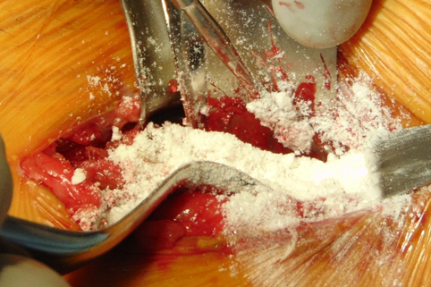
Figure 19. Vancomycin powder is applied over the cement during its hardening process so it can be incorporated to its structure.
After set of the cement, the graft appeared largely stable with no loose fragments or mobility (Figure 20).

Figure 20. After 8 minutes and 30 seconds the hydroxyapatite cement is completely hardened covering the titanium bone plate holes.

Figure 21. Sutures are placed by planes using 3-0 Monocryl® for deep tissues, 4-0 Monocryl® for dermis, and 5-0 Prolene® for epidermis.

Figure 22. Aspect of the final epidermis running suture with 5-0 Prolene® with Mastisol® applied over the area which will receive Steri-strips®, after IobanTM is removed.
Layered closure was performed with sutures 3-0 Monocryl® (Ethicon Inc., J&J, New Brunswick, NJ) for deep tissues and 4-0 Monocryl® (Ethicon Inc., J&J, New Brunswick, NJ), and skin with a continuous suture 5-0 Prolene® (J&J New Brunswick, NJ). Mastisol® (Eloquest Healthcare Inc., Ferndale, MI) Liquid Adhesive and Steri-StripsTM (3M Nexcare, Saint Paul, MN) were applied to support and protect the incision. A total amount of not diluted 10cc of Exparel® (Pacira Pharmaceutical Inc., Parssipany, NJ), a bupivacaine liposome injectable suspension, was given via left IC surgical site. Glasscock® Ear Dressing (Grace Medical, Memphis, TN) was placed along left hip for pressure hemostasis and patient’s comfort (Figures 21, 22).
The total estimated blood loss was 50cc combined with maxillary and mandibular surgical procedures. No complications were appreciated. Patient tolerated the procedure well, was extubated in the operating room and was brought to the recovery room in stable condition. On postoperative days one and two, the patient was seen in his hospital room. He endorsed only minimal discomfort to his hip, but did endorse mild/moderate intraoral discomfort. He was able to ambulate with assistance on postoperative day one. The patient’s pain was managed by use of Exparel injections at the end of the surgical procedure as well as scheduled and as needed pain medications. The patient was maintained on Unasyn throughout his hospital stay to minimize post-operative infection risk. The patient also received dexamethasone 8 mg every hours for the first 24 hour postoperatively. He was able to tolerate a puree diet with adequate caloric intake. Of note, the patient has been treated for the past twenty years for chronic pain with multiple pain medications including methadone. Prior to admission, patient reported having only 1-2 bowel movements per week at baseline. The patient did not have a bowel movement during his postoperative stay in the hospital. This was important due to the surgical procedure having a risk of postoperative iieus.
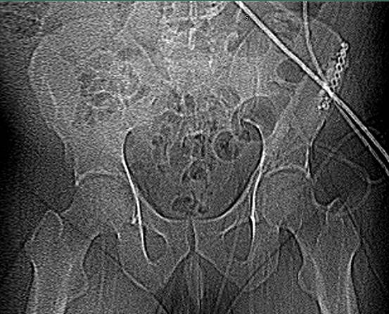
Figure 23. Pelvis x-rays showing titanium bone plate and screws and cement in position.

Figure 24a. First postoperative day Computed Tomography (CT) hip left w/o contract, Coronal view (Cv), showing the most anterior aspect of HA cement adaption, and the initial portion of the Ti bone plate.

Figure 24b. First postoperative day CT hip left w/o contract, Cv, showing the medium aspect of HA cement adaptation, and the head of the second anterior Ti screw.
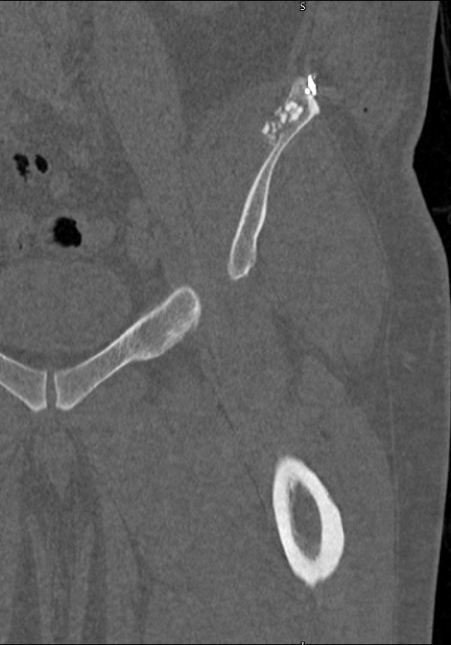
Figure 24c. First postoperative day CT hip left w/o contract, Cv, showing the most posterior aspect of HA cement adaptation, and the head of the second posterior Ti screw.

Figure 25a. First postoperative day CT hip left w/o contract, Sagittal view (Sv), showing the most lateral aspect of HA cement adaption, the initial portion of the Ti bone plate, the head and body of the first and second anterior screws, respectively.

Figure 25b. First postoperative day CT hip left w/o contract, Sv, showing the medium portion of the hip, HA cement adaption, and some mid-portion of the Ti bone plate.

Figure 25c. First postoperative day CT hip left w/o contract, Sv, showing the most medial aspect of HA cement adaption, the head and body of the first and second posterior screws, respectively.

Figure 26a. First postoperative day CT hip left w/o contract, Axial view (Av), showing the most superior aspect of HA cement adaption, and the initial portion of the Ti bone plate.

Figure 26b. First postoperative day CT hip left w/o contract, Av, showing the medium aspect of HA cement adaption, and the mid-portion of the Ti bone plate.

Figure 26c. First postoperative day CT hip left w/o contract, Av, showing the most inferior aspect of HA cement adaption, and the final portion of the Ti bone plate.

Figure 27. Postoperative appearance of the iliac wound on the sixth day, after wound care with Betadine® (Atlantis Consumer Healthcare Inc., Bridgewater - NJ). A large ecchymosis is appreciated posteriorly and laterally to the incision site, as expected, but with no fluids accumulation.
DISCUSSION
Without controversies, the IC remains the most frequently utilized site for autologous bone harvesting when large quantity of cancellous bone is the objective. This donor site has many advantages when compared to other autologous sites such as but not limited to: a) large volume, b) anatomical landmarks which are easily accessible, c) straightforward access from skin to bone, d) better outcomes than other sites, in terms of osteogenesis, e) bone layer beneath the posterior IC does not atrophy so it always can provide enough bone for harvesting.
Complications of IC bone harvesting can include but are not limited to: a) moderate to severe pain, b) nerve injury, c) intraoperative hemorrhage, d) postoperative hematoma formation, e) iliac fracture, f) hernia or intraabdominal injury, g) adynamic ilieus, and h) ureteral injury.
The donor site can require up to three months or even longer to heal and up to 12-16 months for complete histological repair. During the initial healing phase, patients are advised to avoid extreme exercise for up to six months.
Postoperative drains are not usually mandatory for patients undergoing ICBH, but they can be used in certain cases. Now, the use of closed suction drainage in patients undergoing ICBH can significantly reduce the incidence of incision complications, reduce hematoma infection without increasing pain level and reduce hospital stay. However, using a closed suction drainage obviously causes an increase of blood loss, demands drain removal, and produces a visible scar on the skin at the site of insertion.
Various implantation materials and techniques have been used to rebuild iliac defects after bone harvesting. Reconstruction of the IC with use of a hydroxyapatite-calcium triphosphate biphasic compound, which improved the body’s ability to reform new bone but did not alleviate the pain [19].
Other studies have been carried out for the repair of IC defects using autologous bone [15]. The authors of this article do not agree with this concept because it is counterproductive to access another patient’s body area to remove bone to restore the IC when causing another bone defect and the morbidity of a second surgical site which delay the patient’s recovery.
Bovine cancellous grafts, polymethyl methacrylate bone cement, and allografts fixed with cannulated screws have been advocated in 2012 [17], 2013[18], and in 2016 [20], respectively. Although such studies provided some options to reduce donor site pain when trying to reconstruct the IC, the results were not consistent, the material was not homogeneously placed into position and not adequately secured in its final position. Reports of displaced hydroxyapatite cement were described [2].
The authors of this article described a simpler and effective method with better material to easily and anatomically reconstruct patient’s IC after bone harvesting, with full certainty the grafted HABC is firmly secured in position.
HydroSetTM is a self-setting, calcium phosphate cement intended for use in the repair of bone defects as well as in the augmentation or restoration of bony contour in human skeleton. It is formulated to harden even in the presence of water or blood [21,22]. This material after hardened forms hydroxyapatite remodeling to natural bone through osteoclastic resorption and new bone formation. It is specifically designed to set quickly once implanted under normal physiological conditions and it averts thermal injury as it does not give off any potentially damaging heat [23].
These authors agree with a study published in 2009 [24] which recommended that this HABC can be applied manually by hand or spatula or injected through a syringe, enabling better adadption.
IobanTM is an antimicrobial incise drape that is applied to the patient's skin to reduce the risk of surgical site infection. It is known to have several benefits such as: a) creating a sterile surface, b) antimicrobial activity, c) Immobilizing bacteria, d) Conformable adhesion. IobanTM is a Class III medical device which is sterilized using gamma irradiation rather than ethylene oxide, which can produce a skin irritant [25]. Using an iodine-impregnated drape such as IobanTM does not only significantly reduce the resident skin flora, but its use also clearly reduces intraoperative wound contamination, as described in 2010 [26]. Because of these biochemical attributes make this medical device is the authors’ choice for covering the area of surgical removal of ICBH.
The authors suggest that the use a meticulous IC dissection, osteotomies performed with ultrasonic and/or piezosurgery devices, Ti bone plate fixation with Ti bone screws to stabilize pelvis along with HABC, make a significant difference in this type of bone harvesting due to the reconstruction of the anterior IC as reported in 2005 [26]. The authors propose the technique described be considered as an excellent procedure for large amounts of bone collection but a viable and first-line option in terms of pain and local aesthetics. For certain, the use of Exparel® to control postoperative pain is an important adjunct as described before [27], but it does not alleviate ambulation pain and/or discomfort if those previous factors are not taken into consideration for this type of surgery.
General conclusions
Autologous ICBH has been neglected and almost unrecoverable due to several conditions related to bleeding, pain, hematoma, ambulation and visible scar at the donor site. But decreased intraoperative bleeding and postoperative pain as well as improved early ambulation can be obtained through reconstructing the IC defect after autologous harvesting with HABC and corticocancellous Ti bone plates and screws. The technique is simple, safe, and easy to be implemented, making it possible to be performed in most hospitals. However, a larger sample of patients and a prospective, controlled study are necessary to verify and extend our results.
The technique described on this article should be considered an outstanding surgical procedure for ICBH due to its various benefits when compared to the traditional techniques which do not consider donor site reconstruction.
ACKNOWLEDGEMENTS
The authors would like to thank this patient who accepted the treatment and authorized his case description on this publication to revisit the knowledge on ICBH and enhance the techniques for such surgical procedure.
FUNDING
No funding was necessary nor obtained for this case study.
AVAILABILITY OF DATA AND MATERIALS
All data are contained in this paper. The raw data, images in this case report can be available from the corresponding author on reasonable request.
ABBREVIATIONS
Ti: Titanium; IC: Iliac Crest; ICBH: Iliac Crest Bone Harvesting; HABC: Hydroxyapatite Bone Cement; HAC: Hydroxyapatite Cement; CT: Computed Tomography; Cv: Coronal View; Sv: Sagittal View; Av: Axial View.
AUTHORS’ CONTRIBUTIONS
Conception and design: ELLS. Performed the surgery: ELLS, MW, and BB. Design of surgical technique: ELLS. Writing, review, and/or revision of manuscript: ELLS, MW, BB, and PSK. All authors read and approved the final manuscript and its submission for publication.
Consent for publication
Patient participant agreed to publication and signed the consent form.
Competing interests
The authors declare that they have no competing interest, have no association with any medical product company and have nothing to disclosure.
Special thanks
The authors would like to thank Fanar Faeq Subhi, fourth year dental student at WVUSOD, for taking photos and recording videos during the surgical procedure for this case publication.
REFERENCES
- Zenner J, Hitzl W, Mayer M, Koller H. (2015). Analysis of postoperative pain at the anterior iliac crest harvest site: a prospective study of the intraoperative local administration of ropivacaine. Asian Spine J. 9(1):39-46.
- Zhang J, Wei Y, Gong Y et al. (2018). Reconstruction of iliac crest defect after autogenous harvest with bone cement and screws reduces donor site pain. BMC Musculoskeletal Disorders. 19:237.
- Vura N, Rajiv RK, Sudhir R, Rajasekhar G, Kaluvala VR. (2013). Donor site evaluation: anterior iliac crest following secondary alveolar bone grafting. Journal of Clinical and Diagnostic Research. 7(11):2627-2630.
- Ferraz MP. (2023). Bone Grafts in Dental Medicine: An Overview of Autografts, Allografts and Synthetic Materials. Materials (Basel). 16(11):4117.
- Freilich MM, Sándor GK. (2006). In-office iliac crest bone harvesting for peri-implant jaw reconstruction. J Can Dent Assoc. 72(6):543-547.
- Nicolae CL, Pîrvulescu DC, Niculescu AG, Epistatu D, Mihaiescu DE, Antohi AM, et al. (2024). An Up-to-Date Review of Materials Science Advances in Bone Grafting for Oral and Maxillofacial Pathology. Materials (Basel). 17(19):4782.
- Khan SN, Shahzad H. (2023). Osteobiologics and Value-Based Care: Challenges and Opportunities. Int J Spine Surg. 17(S3):S44-S52.
- Willcox MJ. (2016). Lumbar herniation of kidney following iliac crest bone harvest. Case Rep Surg. 2016:5365647.
- Fasolis M, Boffano P, Ramieri G. (2012). Morbidity associated with anterior iliac crest bone graft. Oral Surg Oral Med Oral Pathol Oral Radiol. 114(5):586-591.
- Zermatten P, Wettstein M. (2012). Iliac wing fracture following graft harvesting from the anterior iliac crest: literature review based on a case report. Orthop Traumatol Surg Res. 98(1):114-117.
- Kim DH, Rhim R, Li L, Martha J, Swaim BH, Banco RJ, Jenis LG, Tromanhauser SG. (2009). Prospective study of iliac crest bone graft harvest site pain and morbidity. Spine J. 9(11):886-892.
- Armaghani SJ, Even JL, Zern EK, Braly BA, Kang JD, Devin CJ. (2016). The evaluation of donor site pain after harvest of Tricortical anterior iliac crest bone graft for spinal surgery: a prospective study. Spine (Phila Pa 1976). 41(4):E191-E196.
- Resnick DK. (2005). Reconstruction of anterior iliac crest after bone graft harvest decreases pain: a randomized, controlled clinical trial. Neurosurgery. 57(3):526-529.
- Bapat MR, Chaudhary K, Garg H, Laheri V. (2008). Reconstruction of large iliac crest defects after graft harvest using autologous rib graft: a prospective controlled study. Spine (Phila Pa 1976). 33(23):2570-2575.
- Defino HL, Rodriguez-Fuentes AE. (1999). Reconstruction of anterior iliac crest bone graft donor sites: presentation of a surgical technique. Eur Spine J. 8(6):491-494.
- Chau AM, Xu LL, van der Rijt R, Wong JH, Gragnaniello C, Stanford RE, et al. (2012). Reconstruction versus no reconstruction of iliac crest defects following harvest for spinal fusion: a systematic review: a review. J Neurosurg Spine. 16(6):565-572.
- Makridis KG, Ahmad MA, Kanakaris NK, Fragkakis EM, Giannoudis PV. (2012). Reconstruction of iliac crest with bovine cancellous allograft after bone graft harvest for symphysis pubis arthrodesis. Int Orthop. 36(8):1701-1707.
- Niu YF, An XF, Wu DJ, Xu SG, Zhang CC, Li M. (2013). Anatomical reconstruction of donor site after large iliac crest graft harvest with equivalent iliac crest allograft. A prospective controlled study. Eur Rev Med Pharmacol Sci. 17(14):1951-1957.
- Burton DC, Carlson BB, Johnson PL, Manna BJ, Riazi-Kermani M, Glattes RC, et al. (2013). Backfilling of iliac crest defects with hydroxyapatite-calcium triphosphate biphasic compound: a prospective, randomized computed tomography and patient-based analysis. Spine J. 13(1):54-61.
- Lee JS, Park YJ, Wang L, Chang YS, Shetty GM, Nha KW. (2016). Modified iliac crest reconstruction with bone cement for reduction of donor site pain and morbidity after open wedge high Tibial osteotomy: a prospective study. Knee Surg Relat Res. 28(4):277-282.
- Hannink G, Wolke JGC, Schreurs BW, Buma P. (2007). In Vivo Behavior of a Novel Injectable Calcium Phosphate Cement Compared with Two Other Commercially Available Calcium Phosphate Cements.
- HydroSet IFU. (2025). Available at: https://cmf.stryker.com/assets/files/6v/cmf-br-148_rev.-none_17430-hydroset-brochure.pdf
- Larsson S. (2006). Injectable Phosphate Cements – A Review.
- Clarkin OM, Boyd D, Madigan S, Towler MR. (2009). Comparison of an experimental bone cement with a commercial control, Hydroset. J Mater Sci Mater Med. 20(7):1563-1570.
- 3M Multimedia. Available at: https://multimedia.3m.com/mws/media/2320524O/3m-ioban-2-antimicrobial-incise-drape-interactive-pdf.pdf
- Kramer A, Assadian O, Lademann J. (2010). Prevention of postoperative wound infections by covering the surgical field with iodine-impregnated incision drape (Ioban 2). GMS Krankenhhyg Interdiszip. 5(2):Doc08.
- O'Neill KR, Lockney DT, Bible JE, Crosby CG, Devin CJ. (2014). Bupivacaine for pain reduction after iliac crest bone graft harvest. Orthopedics. 37(5):e428-e434.